Zika – Why Should You Care?

By Sian Venables

Once thought to be a relatively mild mannered virus, Zika’s role in the development of severe foetal abnormalities has put it firmly on the public’s radar. With an epicentre in Brazil it has spread to over 40 countries, upsetting the Olympic games in Rio and causing a national outcry in the US when it required funding to be siphoned from the Affordable Care Act. Hurdles to its eradication arise because of its subtle symptoms and ease of transmission. Whether through targeting mosquitos or developing vaccines, there will be no single solution to quashing the Zika epidemic.  

The WHO recently classed the outbreak as a “public-health emergency of international concern.”. Identified in Uganda in 1947, there had been only 14 documented cases of the virus until 2007, when the first epidemic swept through the Pacific Islands. It cropped up again in 2013 in French Polynesia before the latest outbreak in Brazil was postulated to have links to an infected traveller to the World Cup in Rio, 2014.

Public-health emergency of international concern

The virus itself manifests predominantly in Aedes mosquitos. Though bites from the infected insects are believed to be the commonest route of transmission, spread can also occur through sex, blood transfusions and, to devastating effect, from mother to child. Because the virus can be so harmless, asymptomatic carriers are hard to identify, making upsetting transmission a daunting task. In the French Polynesian out-break, blood banks were thrown into a frenzy; with testing for Zika not previously standard the CDC reported that ‘2.8% of blood donors tested positive’.

Zika’s spectrum of effects is seen in its most extreme form in pregnant women. Infections acquired in the first trimester are associated with the worst outcomes for developing children, so the challenge lies in rapidly identifying these cases. Generally presenting with only a rash, fever, joint pain and conjunctivitis, expectant mothers and hurried health care workers often attribute symptoms of the viral infection to pregnancy hormones.

‘Mounting evidence from many studies’ links maternal Zika infection to the development of severe foetal microcephaly, states the CDC. The virus was identified both in the brains of miscarried micro-cephalic children and in the amniotic fluid of infected mothers (Calvet et al. 2016) It is thought to cause the disorder by proliferating in, and therefore damaging, cortical neural progenitor cells (which go on to form cortex) so is especially damaging to vulnerable, developing brains (Tang et al. 2016).

Though the effects of Zika have mainly been publicised as harmful to developing foetuses, more evidence is building to highlight negative outcomes of infection in children and mature adults. Presence of the virus is now described as a causal factor in the development of the autoimmune disorder Guillain-Barré (Cao-Lormeau et al. 2016). The condition is usually temporary, characterised by the body attacking its own nerves it presents with peripheral muscle wasting and paralysis. Occasionally it affects muscles that control breathing, a consequence that can prove fatal.

Cases of acute inflammation of the brain and spinal cord are also being examined as having a possible link to Zika infection. The development of these conditions means that neurologists will be key players in the fight against the virus, though they are in short supply in some of the worst affected areas.

108% spike in online orders of pregnancy terminating pills

The virus’ spread has given rise to discussion of a taboo ethical dilemma in many of the worst hit countries where abortions are illegal. A case example is Brazil, where abortion is outlawed except in circumstances of rape, health emergencies or anencephaly. Activists are campaigning for laws to be loosened where Zika is a concerned, after an 108% spike in online orders of pregnancy terminating pills in affected areas (Aiken et al. 2016).

This surge in demand for abortions is suspected to be underestimated, as some women in helpless situations resort to unsafe, underground terminations. The task of caring for a severely disabled child is realised as emotionally and often economically unfeasible for many living in already deprived circumstances. The epidemic is not set to die down any time soon, so providing access to safe abortions is an issue that needs to be addressed with an open mind by governments and religious leaders alike, if we are to protect affected women from the social and often fatal consequences of illegal abortions.

With a tried and tested vaccine years from completion, swift reactions to tackling Zika are predominantly focussing on thwarting transmission. Compliance with public health advice about abstinence in pregnancy and avoiding unprotected sex, as well as covering up to avoid mosquito bites at dusk and dawn, are key to keeping infection rates low. A pioneering $18 million project is currently on the go, with intent to release “mutant mosquitos” infected with Wolbachia bacteria into the worst affected areas of Brazil. The bacteria has been reported to transform Aedes aegypti mosquitoes, making them ‘highly resistant’ to two forms of Zika virus, to the extent that they no longer harbour the virus in their saliva (Dutra et al. 2016).

The spread of Zika is likely to be exacerbated by a rise in global temperatures and the unstoppable force of globalisation. With ever more interconnected societies comes a shared responsibility to address the ethical dilemmas and financial burdens of treating infectious diseases. How we handle this outbreak will set a precedent for the inevitable onslaught of epidemics that will come from rising anti-biotic resistance. Zika’s devastating effects can and will be thwarted, but how much pressure is put on governments to treat it as a priority will determine how quickly the battle is won.

If the many dilemmas raised by Zika are of interest to you Medsin urges you to come to a talk hosted by the Hispanic society on the 10th of November:Zika virus: From obscurity to threat’. The event will take place from 6-8pm in School 6 of St Salvator’s quad. We hope to see you there!

Medsin Talk Facebook Event

Bibliography

Aiken, A.R.A. et al., 2016. Requests for Abortion in Latin America Related to Concern about Zika Virus Exposure. New England Journal of Medicine, 375(4), pp.396–398.

Calvet, G. et al., 2016. Detection and sequencing of Zika virus from amniotic fluid of fetuses with microcephaly in Brazil: a case study. The Lancet Infectious Diseases, 16(6), pp.653–660.

Cao-Lormeau, V.-M. et al., 2016. Guillain-Barré Syndrome outbreak associated with Zika virus infection in French Polynesia: a case-control study. The Lancet, 387(10027), pp.1531–1539.

Dutra, H.L.C. et al., 2016. Wolbachia Blocks Currently Circulating Zika Virus Isolates in Brazilian Aedes aegypti Mosquitoes. Cell host & microbe, 19(6), pp.771–4.

Tang, H. et al., 2016. Zika Virus Infects Human Cortical Neural Progenitors and Attenuates Their Growth Brief Report Zika Virus Infects Human Cortical Neural Progenitors and Attenuates Their Growth. Cell Stem Cell, pp.1–4.

Observership Woes

By Ajay Shah

This summer, I spent more than 400 hours in Toronto hospitals. No, not as a patient, but as an unpaid, underappreciated “Medical Observer”. The official role of an observer is limited to simply observing their preceptor, but my responsibilities were vastly different. I would wake up at 5:30 AM and commute an hour to the hospital. If I arrived early enough, I would do morning rounds on my preceptor’s overnight patients. Then, I would rush to the Ortho wing for 6:45 AM morning handover, as the sleep-deprived residents scrolled through X-rays and MRIs, describing unfamiliar and new procedures and fractures. We’d march single file down to the Teaching Room, for 7:00 AM teaching rounds. There, a senior surgeon would speak for an hour about an incredibly specific orthopaedic condition; avascular necrosis of the talar head, anyone? In a valiant effort to stave off sleep, I’d infrequently jot down notes in my notebook, trying to appear engaged. Lest anyone arrive late to these sessions; senior surgeons are ruthless and unforgiving in their admonishments.

Once the morning festivities subsided, the residents rushed off to their respective stations. Pagers were handed over, ER consults were delegated, and patient rounds were polished off. It amazed me how freely people would disrespect colleagues and superiors behind their back; medicine is truly as cutthroat and drama-filled as Grey’s Anatomy would have you believe.

It amazed me how freely people would disrespect colleagues and superiors behind their back

Around 8:00 AM I’d follow my resident into the Surgery wing or Orthopaedic clinic. In surgery, we’d greet the preoperative patients, shave and mark them for surgery, then prepare the operating room. Doctors are so specific about each table, tool and tube; even a slight anomaly can induce profanity-laced tirades. Clean room, patient in, drape & sterilise patient, scrub in, operate, suture, scrub out, undrape, splint, patient out, fill orders, post-op report, clean room, rinse, repeat. Around 3 PM the surgeon would look at the clock, shocked at the slow pace of the day, and blame nurses, porters, residents and students for the slow pace. Finally, around 4:30 PM, the final patient would be bandaged up, sent to post-op wards, and the surgeon would go home. The residents and students were none too lucky. We would be operating in the Trauma or On Call rooms until at least 8:00 PM, seeing post-op patients in between each operation. After a long day operating, we would change out of our scrubs, drive home, and spend the night reading published articles or Orthopaedic textbooks.

Clinic was a different story, although no less stressful. Around 8:00, the triple-booked patients would begin their impatient waits. While we quickly scrolled through X-rays and reports, we could feel the anxiety and resentment from the waiting patients continue to build. Each patient was consulted, examined, and, for the vast majority, told to go home. It is very frustrating to tell a patient, on their third round of referral, that we cannot do anything to help their pain. Even worse is telling a patient that they are not a surgical candidate; their chronic daily pain isn’t severe enough to warrant an operation. Every patient somehow feels marginalised by the system, feels that the doctor is giving them the short end of the stick. Thick accents, learning disabilities, degenerative disorders, and general hatred make some patients difficult to deal with, evoking little of the compassion or empathy we are taught to show in medical school.

So why, one might ask, would anyone want to be a surgical resident. 100+ hour weeks, 48 hour shifts, relentless work, a steady stream of thankless ungrateful patients. Perhaps the emotionally numbing experience is a rite of passage for becoming a senior consultant. Perhaps the long hours build character and motivation, both of which are needed to the utmost when performing operations.

Perhaps the emotionally numbing experience is a rite of passage for becoming a senior consultant

Or perhaps the long hours we spend on rotations, internships and residencies serve to grind us down, to make us bitter and angry. Perhaps experiencing such hard work gives senior surgeons a sense of entitlement, and a licence to treat their junior colleagues poorly. Perhaps this poor treatment propagates a vicious cycle of unfriendliness, burnout, and resentment. Maybe the general malice is a quality necessary in brilliant surgeons; their meticulousness and attention to detail may irk some people, but it certainly gives good outcomes.

I learned a lot this summer about medicine. But I learned more about people. I saw amazing residents, and I saw incompetent residents. I saw funny, friendly surgeons, and I observed arrogant, crusty old surgeons. These articles usually end with a parting piece of advice, so mine will be this:

Don’t be afraid to kiss ass, laugh at unfunny jokes, or go the extra mile. Show up early, don’t make mistakes, and be the last to leave. Those who work the hardest now will reap the most reward when the time comes. Take time to educate and mentor those younger than you, and be friendly to everyone you meet. Wisdom is hidden behind clichés.

An Oxford University Press Book Review

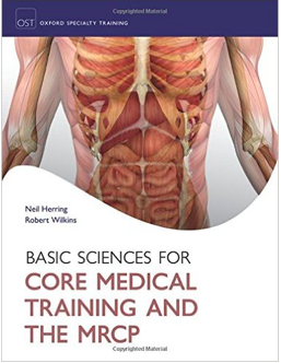
Both Oxford Cases in Medicine and Surgery and Basic Science for Core Medical Training are newly published titles having come out just this past year. They promise to be useful additions to your medical textbook collection that contain essential knowledge for medical students transitioning from purely academic knowledge and needing to apply this knowledge clinically. Plus, they are concise and straight to the point, which means you learn what you need to be ready for a clinical environment and no heavy lifting.


Basic Sciences for Core Medical Training and the MRCP

Oxford University Press, Edited by Neil Herring and Robert Wilkins

Fresh off the OxUntitledford university press, comes a new textbook designed specifically, or so it seems, for the pre-clinical St Andrews student. Using the same systems-based approach, with integrated clinical relevance that our curriculum throws at us, this brand new book clearly lays out all the basic science necessary for the typical honors student. But how does the book really perform next to the average recommended medical booklist?
With plenty of information to cram into your head, and what seems like too few hours in a day, organisation is key in the life of a medical student. So the first thing you may notice about this book is how well it’s laid out. Covering all the major topics, from cell biology to endocrinology, the book moves logically through the systems and specialties, covering all the basic knowledge you could ask for, in as simple terms as possible.


If you’re an auditory learner, the clear layout of information, with bullet-pointed summaries will be the perfect resource for you… lists, tables and charts are used to summarise each chapter concisely into about 1 A4 page. However, there are plenty of illustrations too; well-labeled diagrams are scattered throughout. The one disappointment however is the lack of colour. It can be difficult to see the detail, particularly in the histological images (which are only coloured in black, white and purple), so honestly, I’d recommend looking elsewhere if you want to see some clear microscopic imagery. But then again, the book does make a fantastic information reference point to keep open next to you while you’re looking.


The flow chart summaries are particularly helpful when looking for a quick revision tool. Stuck up on your wall or on the back of your door, these illustrations work perfectly as memory prompters to test yourself, especially when it comes to the complicated, intertwining pathways of cardiac physiology.


System and subject specific testing is made easy, as each chapter ends with a set of practice MCQ’s. The questions have a familiar format; a pre-amble with a clinical scenario, some key basic science knowledge thrown in and then it’s time to diagnose or treat the patient. The only problem, is quite frankly, there aren’t enough of them! With only 10 MCQ’s at the end of each chapter, only a minuscule percentage of the content is actually touched upon in the questions.


In fact, this is the main drawback of the books entire content… there just isn’t enough. The “basic” knowledge really is what this book provides. The clear and concise information is really only the foundation of what is covered over the 3 pre-clinical years here in St Andrews. So although the perfect starting point for revision, don’t throw away that book list with delight… it’s clear that it’s only one resource of many, and for the real detail, you will need to supplement with additional resources.


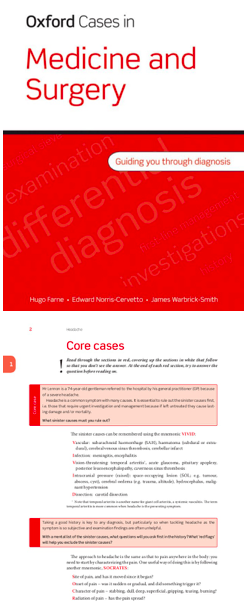
Oxford Cases in Medicine and Surgery

Oxford University Press, Hugo Farne, Edward Norris-Cervetto, and James Warbrick-Smith

Untitled 2Oxford Cases in Medicine and Surgery promises to be unique in that it’s goal is essentially to teach students how to think clinically without needing to be in a clinical setting. Different, but very common presenting complaints are the focus of each chapter, or ‘Core Case’ as they have been titled. Each Core Case begins by stating the name, age and presenting complaint of a patient and then, based solely on that information, asks you to think critically and identify all possible serious causes that you need to rule out first. Sound like something every medical student needs to know how to do when they get out on the wards? Definitely. However, instead of throwing you in the deep end filled with anxiety, as you would feel on a ward with an actual patient, Oxford Cases gives you mnemonics and walks you through all of the red flags you should be looking out for. This text teaches you how to evaluate patients’ presenting complaints in a systematic way that ensures you won’t be missing anything vital and are covering all possibilities efficiently.


 

In addition, each time you eliminate a possible cause in a core case, Oxford Cases pushes you further, continuously asking you why to broaden your understanding and getting you ready for the real thing. These informal questions scattered throughout the Core Case are supplemented by ‘Viva’ questions and SBAs (Single Best Answer questions) at the end of each chapter. The Viva questions are a set of questions meant to test your overall understanding of what was just taught in the Core Case in addition to some ethical dilemmas you may face. The SBAs are designed to have you master multiple-choice questions on your medical papers in no time.


Within each Core Case, ‘Short Cases’ are also presented which briefly explain another clinical case of a patient with the same presenting complaint, but ultimately has a different diagnosis. This, again, pushes you to think clinically and challenges you to draw from your previous academic knowledge and apply it. At the end of each ‘Short Cases’ section, Oxford Cases clearly site which research papers were used so, if you’re extra keen, you can easily find them to read more detailed information.

How to Get Published and Be Successful

By Ajay Shah

12833278_899794723449469_1397449614_n

2nd year medical student Ajay Shah, writes about the anxiety surrounding lab research and publications- something all medical students can relate to!


“Have you done any research?”

             “Yeah actually I have a publication in the Canadian Journal of Obstetrics, I spent my summer doing a meta-analysis for Mount Sinai”

“Oh, so do you want to do obstetrics?”
               “No, I just managed to get the position through a friend. I don’t actually care about it, research is kind of boring” 


I’ve had this conversation with so many students, at St. Andrews and otherwise. It seems to be hammered into us from an early age. “Get published, get hired” is the prime directive for stethoscope-wielding youth. For Canadian students, the pressure is multiplied, as the prestigious residency positions will only accept the most credentialed, cited and publication-producing students.

____________________________________________________________


So, we slave. I am no exception to this rule, I write from my experiences. In high school, I spent most of a year doing a research project at Princess Margaret Hospital. Today, if you saw me standing with my poster at conferences, you’d be smitten by my passionate descriptions of MTS Assays and In silico analysis. If you saw me back then, in that stuffy lab from 5-7 PM everyday, micropipetting row upon row, redoing protocol due to calculation errors, waiting for colonies to incubate, you’d feel nothing but pity.

12804496_899794720116136_1558064817_n

‘you’d feel nothing but pity’

And that is the reality of lab research. It is boring. You will fail. Even when you do everything right, you will get negative results, contamination, and false positives. My project looked at the role of microRNAs in human breast cancer cells, and we received negative results with the first two microRNAs we tested. After 8 months in the lab, we concluded that microRNA-449 levels were correlated with tumour viability, then found 4 genes that potentially contribute to tumourigenesis. Peanuts, in the big picture – but another tree planted in the forest of cancer research.

-‘Even when you do everything right, you will get negative results, contamination, and false positives’-

I am not trivializing lab research, nor am I insinuating that incremental accomplishments are meaningless. I can imagine nothing more fulfilling than conducting scientific inquiry into one’s passions. I am simply trying to relate a feeling I had to one that many of you may experience, the feeling of purposeless disappointment. The infinite thought loop of “What exactly have I accomplished?” to “Was that a worthwhile use of my time?” to “What did I gain from that?”, rinse and repeat.

I believe that feeling arose because I was doing research for all the wrong reasons. I was doing that project to get my name on a paper to improve a university application to increase my odds of getting a slightly better education which would, theoretically, ultimately help me achieve the abstract notion of “success”. The idea of success instilled in us by our superiors, mentors and role models, a concept I could hardly visualize, let alone materialize. My search for material scientific findings had an ulterior purpose, and my findings therefore seemed purposeless and immaterial.

-‘that feeling arose because I was doing research for all the wrong reasons’-

So to all those go-getters doing research this summer: I beg you, be wary. If you lack passion in your topic, and are simply doing research to get published, think twice. Perhaps an unpaid shadowing position may help you learn more, think more, and gain a sense of purpose. Or perhaps this mental purgatory is a necessity that we medical students must pass through on our journey – jumping through hoops, dotting I’s and crossing T’s, going through the motions, as we wait for our careers to begin. Whatever it may be, I suppose it can’t hurt to ask – “Hey, do you know anyone hiring students to work in a research lab?”

____________________________________________________________

If you are interested in a particular medically-related topic, please submit your article to medsaint1@gmail.com. As per usual, there is no deadline!


Restless Leg Syndrome: A Brief Overview

12669205_882470808515194_69336108_o

3rd year Caroline Cristofaro investigates restless leg syndrome, a relatively unknown condition that can significantly impact a patient’s qualIty of life.


Introduction:

At first restless leg syndrome may seem like an innocent condition, however, it can be very debilitating for the patient. In addition to the obvious symptom of the patient feeling the uncontrollable desire to move their legs, patients can often suffer from insomnia, parasthesias and various uncomfortable sensations in the lower limbs (Facheris, M., et al., 2010). The goal of this article is to shed light on the symptoms and aetiology of this little known syndrome and what medications are given to treat it.


Causes:

RLS has been found to have a strong genetic link and most cases are diagnosed as Familial RLS. Secondary RLS can be experienced during pregnancy and renal failure, both of which are associated with iron deficiency (Facheris, M., et al., 2010). Opioid-dependent patients can also experience RLS after stopping opioids as a withdrawal symptom (Ghosh, A. et al., 2014).


Symptoms:

Restless Leg Syndrome (RLS) is categorized as both a sensorimotor and sleep disorder with the sensorimotor symptoms being the main complaint in most patients. The sensation is described using a myriad of terms such as itching, burning, crawling, tingling but is relived, albeit only temporarily by movement of the legs (Facheris, M., et al., 2010). Depending on the severity of the patient’s RLS, relief can be by simply stretching out the affected leg(s) or the patient may have to get up and walk around. Although it is fairly obvious why RLS would delay the onset of sleep, the explanation as to why RLS causes disturbed and fragmented sleep is somewhat less evident. Periodic Limb Movements of Sleep (PLMS) are defined as “semirhythmic movements of the limbs which last a few seconds and occur at regular intervals” (Cotter, P. and O’Keeffe, S., 2006), as is indicated by their name. These limb movements are nearly always present in the population suffering from RLS and, in addition, these movements are typically exaggerated and cause patients to awaken leading to patients’ awareness of RLS sensorimotor symptoms, which, of course, makes the task of falling asleep more arduous (Cotter, P. and O’Keeffe, S., 2006).

11982830_864747250288882_327197523_n.gif


Pathophysiology:

Whilst the exact cause of RLS has not yet been cemented, two factors have been shown to heavily impact symptoms. These are iron and dopamine levels within the substantia nigra.

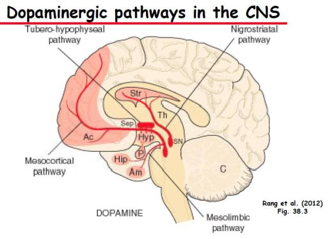
Dopamine is a neurotransmitter, which plays an important role in regulating movement. The two main receptors for dopamine in the brain are D1 and D2 receptors and, each via a specific pathway, increase or decrease movement, respectively. In RLS, Positron Emission Tomography (PET) scans have consistently shown decreased activity of the D2 receptor, which normally decreases movement, leading to increased movement (Ruottinen, H.M., 2000). In support of these findings, Connor, JR et al. (2009) demonstrated a 30% reduction in D2 receptors in the basal ganglia using autopsies of patients who had been affected with severe RLS. As RLS occurs in the lower limbs, it is thought that the dopaminergic A11 cell group is mainly affected because they are the only neurones which provide dopaminergic axons to the spinal cord. This hypothesis was strengthened by Clement, S. et al. (2006) demonstrating increased movement in mice with a lesioned A11 dopaminergic cell group.

12666484_882470801848528_1717728918_n

Iron acts as a coenzyme for tyrosine hydroxylase (an enzyme essential in dopamine synthesis) and it is also related to monoamine oxidase (an enzyme which degrades dopamine) (Facheris, M., et al., 2010). Due to its close relationship with dopamine, the concentration of iron in the brain influences the numbers of dopamine transporters and dopamine receptors (specifically D2 and D4); decreased concentration of iron in the CSF leads to decreased densities of dopamine transporters and receptors which have been attributed to the cause RLS symptoms (Omer, P. et al, 2007). However, a recent study by Conner J.R., et al. (2009) compared the post-mortem tyrosine hydroxylase concentrations of patients who had suffered from RLS to a control group and found no significant difference. Due to conflicting evidence, the previously explained role of dopamine in RLS is considered to be a hypothesis and, as a result, is still referred to as the “Dopamine Hypothesis”.


Treatment:

A critical review conducted by the International Restless Legs Syndrome Study Group in 2013 investigated the efficacy of various drugs to treat long-term RLS. For first line treatment it is recommended to give the patient dopamine-receptor agonists or calcium channel antagonists (pregbalin or gapapentin encarbil) (Gracia-Borreguero, D., et al., 2013). The dopamine agonists recommended are either ergot-based (pergolide and cabergoline) or non-ergot based (pramiprexole, ropinirole, rotigotine, levodopa). Calcium channel antagonists are used mainly when the patient complains of the physical symptoms and act to stop excessive muscle contraction. Iron supplements are also recommended when the patient’s serum ferritin levels are low (Gracia-Borreguero, D., et al., 2013). Together, these treatments decrease muscle contraction, help stabilize the dopaminergic pathways in the brain and increase the patient’s serum iron levels, further stabilizing the concentration of dopamine.


Conclusion:

Although RLS may seem like a relatively benign condition at first thought, its constant, intrusive symptoms encroach on nearly every aspect of a patient’s life, making it very difficult to live with. Both its pathophysiology and, consequently, its treatment options draw similarity to Parkinson’s disease, which should also alert you to the seriousness of this syndrome. Unfortunately, none the drugs used to treat the condition have been proven to be effective for more than 1 year (Gracia-Borreguero, D., et al., 2013), which means that we have yet to find a drug that can effectively treat RLS for when the syndrome persists over a year. Hopefully future studies are able to find a concrete link between dopamine and RLS to explain the efficacy of dopamine agonists in treating the condition, or otherwise determine the accurate aetiology of the syndrome to prompt studies of new drugs able to effectively cure RLS.


Bibliography:


Clemens, S., Rye, D., & Hochman, S. (2006). Restless Legs Syndrome: Revisiting the Dopamine Hypothesis from the Spinal Cord Perspective. Neurology, 67(1), 125–130.


Cotter, P. E., & O’Keeffe, S. T. (2006). Restless Leg Syndrome: Is it a Real Problem? Therapeutics And Clinical Risk Management, 2(4), 465–475.

Garcia-Borreguero, D., Kohnen, R., Silber, M. H., Winkelman, J. W., Earley, C. J., Högl, B., … Allen, R. P. (2013). The long-term treatment of restless legs syndrome/Willis–Ekbom disease: evidence-based guidelines and clinical consensus best practice guidance: a report from the International Restless Legs Syndrome Study Group. Sleep Medicine, 14(7), 675–684.


Ghosh, A., & Basu, D. (2014). Restless Legs Syndrome in Opioid Dependent Patients. Indian Journal Of Psychological Medicine, 36(1), 85–87.

Oner, P., Dirik, E. B., Taner, Y., Caykoylu, A., & Anlar, O. (2007). Association Between Low Serum Ferritin and Restless Legs Syndrome in Patients with Attention Deficit Hyperactivity Disorder. Tohoku J. Exp. Med. The Tohoku Journal Of Experimental Medicine, 213(3), 269–276.


Pramstaller, P. (2010). Update on the Management of Restless Legs Syndrome: Existing and Emerging Treatment Options. Nature And Science of Sleep, 2, 199–212.


Rios Romenets, S., & Postuma, R. B. (2013). Restless Legs Syndrome. Current Treatment Options In Neurology, 15(4), 396–409.


Winkelman, J. W. (2006). Considering the Causes of RLS. European Journal Of Neurology, 13, 8–14.

OMG Bacon Causes Cancer! (When Pigs Fly…)

By: Ajay Shah (2nd Year)

12477218_864821473613461_1407002002_o

In our first article of 2016, Ajay Shah writes about his experience with the news headlines’ exaggeration of research paper results and discovering how to interpret them.


I.

On October 26th, 2015, the results of a new study rippled across social media and news outlets. Sensationalist Twitter hashtags and Facebook posts lamenting its results dominated the cyber landscape. Sales at supermarkets and grocery stores fell by millions of pounds in the following weeks. Thousands of people swore off their favourite foods in the panic, hoping to bargain for a few extra years of life. What could this study have discovered to cause such a viral reaction throughout society? Well, of course, it showed that bacon causes cancer!


Hold up. What? One of the most universally loved foods in the world (Americans consume 18 pounds per capita annually), cast aside by a suddenly health-conscious population, just because of one study’s results? How could this have happened? As is often the case, the culprit is not those responsible for creating the study (they just did the science), nor the general population (we just read the articles). The culprit is far more insidious, something capable of unnecessarily inspiring fear into millions of innocent people. That culprit was The Headlines.


II.

I woke up that morning to see a short BBC notification on my phone declaring that red meat causes cancer. Confused, I called my girlfriend as I skimmed the article.


“WHAT??” said my girlfriend when I read the headline to her, likely picturing morsels of steak morphing into tumours as they passed through her colon.

“Yup, the WHO has declared that red meats cause colon cancer. It’s true – even the BBC is reporting it,” I replied.


After some quick research, and reading headlines and tweets such as:
“OMG! Bacon causes cancer!” (New York Post)

“Processed meats rank alongside smoking as cancer causes — W.H.O. U.N. health body says bacon, sausages and ham among most carcinogenic substances along with cigarettes, alcohol, asbestos and arsenic.” (The Guardian)

“Bacon, hot dogs and processed meats cause cancer/are as dangerous as smoking, says @WHO.” (PBS)


the alarm bells began to ring. My girlfriend quickly swore off processed meats for life, and limited red meats to one meal per week. I, an avid meat-eater myself, decided not to let The Headlines fool me, and began to do some research of my own.


12459955_864821466946795_770154967_n

III.

As I had suspected, the initial reaction to the study was quite overblown. In fact, the first paragraph of the WHO report itself says that “red meat is probably carcinogenic to humans based on limited evidence ”. “Limited evidence” means that, while a positive correlation is present between the exposure (meat) and outcome (cancer), other explanations (confounding factors, bias, chance) cannot be ruled out.


The findings are based on an assessment of more than 800 studies, which gave more weight to the studies with larger sample sizes, fewer confounding factors and better experimental design. Approximately two thirds of the case-control/cohort studies showed a positive correlation between eating processed meats and colorectal cancer incidence. It was found that eating 100g of red meat per day raises colorectal cancer incidence by 17%, while eating 50g of processed meat daily raises incidence by 18%. Both types of meat consumption were also linked to pancreatic, prostate and stomach cancer.


Personally, I think that the study was very well-conducted and is a fantastic example of “Good Science”. The researchers importantly identified a mechanism of action, identifying known carcinogens produced during the cooking of meat, and postulating that these compounds lead to cancer formation. They also examined a wide range of studies under specific criteria, minimizing the influence of confounding factors and bias. The different types of meats were classified appropriately, and suitable recommendations were made.


IV.

Upon reading the WHO report, I felt quite upset at the media for their treatment of the story. The WHO researchers were quick to reassure, suggesting that a reduction of processed meat consumption would be a preferable alternative to widespread vegetarianism. Indeed, they identified the valuable nutrients and compounds in red meat, and after contrasting these with the increased cancer risks, officially only recommended cutting down red meat consumption to 70g daily. This is a stark contrast to the doom-and-gloom picture portrayed by the media.


An increase of cancer incidence by 17-18% may seem like a lot, and with the media’s handling of this report, any meat eater would do a double-take before chowing into that cheeseburger. However, this “relative risk” looks a lot different when put into perspective. It is estimated that, among 1000 people who eat little-to-no processed meat, 56 would develop bowel cancer during their lives. Comparatively, of the 1000 people who eat the most processed meat, 66 are expected to develop bowel cancer during their lives. Indeed, the 17% increase seems miniscule when compared to smoking (2,400% increased risk) or drinking alcohol (500% increase). And this ignores the fact that red meat offers many healthy nutrients, which may overpower the carcinogenic effect in some. Regardless, red and processed meat now joins an increasingly cluttered list of carcinogens, including aloe vera (Class 2B), the Sun (Class 1) and Air (Class 1), some of most dangerous things known to man.


V.

So, throughout the journey from “my bowels are full of carcinogen-induced tumours” to “eating meat probably, might, slightly increase the risk of developing a tumour in my large intestine”, I learned a lot about clinical studies, the media, and The Headlines. I learned that “carcinogen” is a broad term, and a subatomic particle that may be essential to the fabric of our reality (neutrons) is also a known human killer. I learned that clinical studies completed under similar conditions could have polar opposite results. But most importantly, I learned that in a world dominated by clickbait headlines and diminishing attention spans, the onus is on us to do our research, and Rewrite the Headlines.


  1. 955.

Looking for Writers and a Publicity/Social Media Representative!

Hello Everyone!

MedSaint is the University of St Andrews’s medical journal and we’re looking for people to get involved. We’re hoping to gather more writers who are keen to put articles forward to be published on our website. You would be free to choose whatever topic related to medicine that interests you and, best of all, we have no deadline but publish we the pieces as soon as they’re ready. Have a look at our previously published articles on our website to get an idea of what’s been included so far. We’re also looking for someone to fulfill the role of publicity/social media representative for MedSaint. If you’re interested in this position, please send a paragraph of 150 words max to medsaint1@gmail.com telling us why you think you’re suited for the role. Even if you aren’t a medic, but are interested in writing on current medical events, please do get in touch as this isn’t solely for medics.

Hope to hear from some of you soon!

Caroline Cristofaro and Lauren Wong

MedSaint Editors

Eureka? A book review of a brand-new series of medical textbooks

Eureka is a series of textbooks freshly published in 2015 that claims to be specially designed for the modern-day medical student, relevant across all of his or her years of study. Consider this proposition within the context of today’s educational environment, which is populated by universally accessible handouts, online multimedia resources, eBooks, and collaborative Web 2.0 platforms such as ‘Meducation’, ‘MosaicED’, ‘GeekyMedics’, ‘Instant Anatomy’ and ‘TeachMeAnatomy’. With information available and openly accessible, is there an important role marked out for the textbook, and if so, how does Eureka fare as a supplement?

In a textbook, the medical student seeks guidance, companionship, essential content, and clarity of concept.

The discipline of medicine is of long tradition and application, and for this reason, learning and teaching will necessarily drive much of our activity as healthcare professionals. Whilst we appreciate that these are lifelong processes, there is a certain corpus of biomedical knowledge that must be mastered by the medical student before he or she is deemed competent to practice safely on patients. For many of us, it can be difficult to ascertain where to begin in this learning process.

The textbook fills this gap by providing relevant information for immediate reference; a reader need not seek information in a piecemeal manner. Typically, textbooks are divided by scientific discipline; we use the stalwarts Grey’s for Anatomy, Kumar & Clark’s for Clinical Medicine, or Rang & Dale’s for Pharmacology. This canon drips in extraneous detail, and does not directly complement the medical curriculum we are examined for.

Eureka‘s title is immediately captivating, as it appears to imply a solution. Speaking from the viewpoint of pure aesthetics, its covers are simple, elegant and non-distracting; the corners of its pages are beautifully curved; the books themselves are light and easily portable. The books are not overly hefty! As medical students, we have become used to toting around heavy textbooks to the detriment of our backs. The fact that these books are not unnecessarily large or heavy is definitely a bonus, but the question needs to be asked – is the content sufficient, or compromised? 

Each book integrates a system’s core biomedical science with its clinical
– medical and surgical – applications. These books are complementary of the tendency of medical teaching towards a ‘systems-based approach’.

  • What precisely does this ‘integration’ entail? Each book is divided into a set of chapters, which invariably include ‘First principles’ and ‘Clinical essentials’, that cover the anatomy, embryology, and physiology of the system in question, as well as the principles of clinical examination, investigation, the observation of basic signs and their clinical correlates. For ‘Cardiovascular Medicine’, for example, this would entail the structure and function of the heart, the cardiac myocyte and its electrophysiology, cardiac embryology, the arterial supply to the body, and cardiac physiology. This would be followed by examination and interpretation of cardiovascular signs, interpretation of the ECG and the echocardiogram, the cardiac MRI and angiography, amongst the interpretation of results from other modalities. The books do not provide a step-by-step guide to clinical work, but certainly, a more than adequate starting point to its theoretical basis.
  • In this sense, the first chapters cover what one might typically consider the ‘preclinical’ necessities, and do so in sufficient detail, which is an impressive achievement for one volume. The graphics and illustrations are clearly drawn, and these are interspersed with images of radiographs, gross pathology, and clinical photographs, where relevant. It must be noted that the anatomy is not covered in the depth, or captured through the multiple viewpoints and layers, a book such as Gray’s Anatomy would – but then, Eureka does not brand itself as a specialist’s handbook. In a similar vein, it would be foolish to expect annotated histological specimens, or diagrams detailing the specifics of molecular biology. Ultimately, this is not a book designed for the surgeon, pathologist, or molecular biologist, but for the medical student, for whom it is incredibly useful, both clinically and otherwise.
  • In this sense, the first chapters cover what one might typically consider the ‘preclinical’ necessities, and do so in sufficient detail, which is an impressive feat. A book like Gray’s Anatomy can often overwhelm students with technical terms and heavy content. The ‘first principles’ section of the Eureka series assumes that the student most likely already has knowledge of the subject, and its main purpose is to consolidate and summarise that knowledge. It does not go into substantial amounts of detail for anatomy and physiology, but it is a good level of information for a medical student with some prior learning in the area. Particularly for students in clinical years, it is a good refresher of information that may have been learned several years ago. In this respect, the first principles section is an excellent summary of the topic.

The initial storytelling is succeeded by several chapters detailing the most common pathology, where the standard of clear, smooth-flowing, writing, is upheld. These entail the epidemiology, aetiology, pathogenesis, clinical features, diagnostic approach, and medical and surgical management, for a broad range of commonly encountered conditions – as well as a chapter dedicated to emergencies. A public health approach is integrated; for instance, the diagnosis of respiratory disease is intimately linked with a patient’s occupational history, and this is both appreciated and expounded upon in ‘Respiratory Medicine’. Pharmacological interventions are also covered, if not to a depth where their molecular mechanisms are elucidated. Clinical cases are routinely incorporated, and sometimes through realistic ‘graphic narratives’, which are enjoyable to trace. Each chapter is prefaced by a number of thought-provoking questions, that if paid attention to, prompt the reader to actively seek answers – such as ‘What investigations are useful for someone presenting with chronic dyspnoea?’ or ‘What factors determine the surgical approach in a woman shown to have breast cancer?’

As everything from presentation, epidemiology, aetiology, clinical signs, diagnostics and imaging are discussed, this is particularly useful for medical students who feel lost when applying knowledge to a clinical case. By presenting common cases and clearly laying out the steps one would need to take, the information is very approachable and understandable. Boxes are also used to highlight important information, and diagrams are clear and easy to follow. While knowing basic scientific knowledge is certainly important, without an understanding of how it applies to clinical scenarios, it is almost irrelevant! In this manner, the Eureka series does a wonderful job of demonstrating clinical applicability.

The series is edited by Janine Henderson, David Oliviera, and Stephen Parker, who are consultants and teachers in their respective fields and institutions, with two of the authors marking out their personal interests in medical education methods. Each textbook is written and edited by three or four consultants on their respective field, individuals who have invariably had research and teaching experience. This is reflected in the writing, which never confuses, obfuscates, or leaves loose ends for the reader – parentheses are often used to enclose or define technical terminology.

To recap, it would be correct to point out that the systems do not operate in isolation, and certainly, this nuance is appreciated in each textbook – which covers each system holistically. As medical courses in Britain are typically separated into systems-based ‘blocks’, I would highly recommend Eureka‘s textbooks as referential and revision resources for those who consider a portable, efficient, and all-inclusive guide a worthwhile investment. While the Eureka books are a new and emerging series on the market, they definitely prove their worth. The ability to condense information into a relatively small book while still retaining essential content is not an easy feat. Extensive clinical relevance is another great aspect of the books. In short, we would highly recommend the series for medical students looking for a way to consolidate their knowledge in a straightforward manner.

The series, at present, is available at www.eurekamedicine.co.uk. We are very happy to offer a discount code (MED15) for the books when ordered for the website, which will entitle readers to a 15% discount plus free delivery. The offer is available until 31.12.15. All are priced at £22.95 each; with the discount they are £19.50 each. The following titles are currently available:

  • Physiology
  • Biochemistry & Metabolism
  • Cardiovascular Medicine
  • Respiratory Medicine
  • Endocrinology
  • General Surgery & Urology

The following titles will be available from the specified dates:

  • Neurology & Neurosurgery (September 2015)
  • Psychiatry (September 2015)
  • Obstetrics & Gynaecology (January 2016)
  • Gastrointestinal Medicine (January 2016)
  • Renal Medicine (January 2016)
  • Paediatrics (July 2016)

These textbooks were reviewed by Aksha Ramaesh, Angela Hu, and Anahita Sharma, third-year preclinical medical students based in St Andrews, continuing their clinical studies at the University of Edinburgh, McMaster University, and University of Manchester, respectively. The authors have no conflicts of interest to declare.

Prosopagnosia – Lost in a sea of faces?

Screenshot 2015-04-01 21.39.39Jackie Liu discusses prosopagnosia, a cognitive disorder that leaves sufferers struggling to recognise the faces and identities of those around them.


Prosopagnosia is that awkward and embarrassing encounter where you are greeted by someone you supposedly know, but whose face you simply cannot recognise. It’s September in St. Andrews, you meet hundreds of new students, but a few days later in the street you have no recollection of meeting this individual because you cannot recognise their face. For some, this is a way of life, a daily threat, a constant unease. Imagine being in a crowd at a football match, lost and alone, not recognising anyone: not a single identifiable face. Prosopagnosics face this possibility everyday. This condition can lead to severe consequences.

What is prosopagnosia?

Prosopagnosia, or ‘Face Blindness’, is defined as having an inability to, or difficulty with, recognising faces: a cognitive disorder of facial perception. Some cannot perceive faces, and others cannot relate the face to an identity. This, for most people, is a simple and at least a partially unconscious process developed soon after birth, but in some this cognitive process is impaired or missing. The German psychologist Joachim Bodamer first used the term prosopagnosia in 1947 in his paper titled ‘Die Prosop-Agnosie’. It was a derivation from the classical Greek “πρόσωπον” (prósōpon), meaning ‘face’, and “αγνωσία” (agnōsía) ‘non-knowledge’ (Bodamer, 1947).

How does facial recognition work?

To understand prosopagnosia, it is necessary to understand the development of facial recognition. The process is complicated, and seems to begin in babies soon after birth. Newborn infants have been observed to show interest in, and track, basic sketched faces. This innate interest declines after the first month. This development is crucial to facial recognition as researched by Grand et al. (2001). The study examined patients deprived of normal vision with bilateral congenital cataracts, which were surgically corrected 118 days post-birth on average. This delay, however, caused an insufficient development of the facial processing ability. These patients were compared to a control group who had had no previous visual-impairment and the results indicated that deprivation of facial visual input at birth led to permanent deficits in configural facial processing – the spacing of facial features. Yet the visually deprived patients had normal featural processing – the shaping of features. So while these patients can process and recognise the shapes of features like eyes or nose, their deprivation of visual input at birth may have led to a permanent difficulty in recognising differences in the spacing of features, and hence the inability to connect identity with a specific set of facial features (Grand et al., 2001). Prosopagnosia is a defect specifically with the holistic or configurational processing of faces (Busigny et al., 2010).

Hence, prosopagnosia may lead to the dissociation of two functional processes: the recognition of a face through its’ features, and spacing. Spacing is a function that is separate from featural processing, but assists in the identification of the owner of the face, which has been discussed as a secondary process by Schiltz (2005). This paper suggests that there are indeed two levels of processing: face detection, followed by individual identification, and that these levels can be dissociated (Schiltz, 2005). In a person without prosopagnosia, these processes integrate, working largely unconsciously to enable facial recognition.

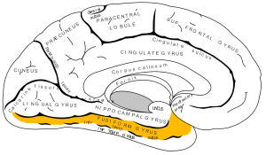
Anatomically, there is debate (Halgren et al., 2000) with regards to the location of facial recognition processes, but the general consensus is that it functionally localises to the occipitotemporal or fusiform gyrus.

Occipitotemporal or Fusiform Gyrus

Within the gyrus is an area called the ‘Fusiform Face Area’, which is suspected to function in facial recognition (Kanwisher & Yovel, 2006). Functional MRI (fMRI) reveals an increase in blood flow in the Fusiform Face Area when the patient looks at faces. The evidence shows that the Area demonstrates functional specificity in relation to faces rather than objects, and also area specificity suggested by the differences in response profiles from other face-selective regions (Kanwisher & Yovel, 2006). However, some argue (Goldstein, 2009) that the area cannot be the sole anatomical basis for facial recognition, as it is not fully developed until adolescence, and babies can differentiate faces as early as 3 months old. It also argued that cognitive functions, like facial recognition, are not limited to being domain-specific mechanisms (Kanwisher & Yovel, 2006).

Is there a difference between the two hemispheres of the brain?

Studies have suggested that the facial recognition process is expressed from the right hemisphere (Meadows, 1974). Patients with acquired prosopagnosia develop the condition after traumatic brain injury or damage. Meadows (1974) describes prosopagnosia patients as almost always having a right occipitotemporal lesion (Meadows, 1974). A study by Schiltz (2006) also provides supporting evidence for this theory, indicating a correlation between lesions in the fusiform gyrus on the right hemisphere with difficulties identifying faces (Schiltz, 2006).


What are the different types of prosopagnosia?

Prosopagnosia is not a unitary disorder: there are different types and levels of impairment. The condition can be categorised into two main groups: acquired and developmental. Acquired has two sub-groups:

  • Apperceptive prosopagnosia is impairment to the earliest processes in facial recognition, usually caused by damage to the right occipitotemporal region. These patients may be unable to recognise faces at all, whether familiar or not. These patients are then dependent on other factors, like voice and clothing, for recognition (Gainotti & Marra, 2011). 
  • Associative prosopagnosia is impairment to other early face recognition processes and also their links with memory, usually caused by damage to the right anterior temporal regions. These patients can determine faces and what the face may show about the person (age and sex), but cannot associate the face with anyone they know; there is no identification (Gainotti & Marra, 2011).

Developmental is defined as being present from birth, and manifests in early childhood (Jones & Tranel, 2001). Inheritance is possible: there are families with multiple members affected by prosopagnosia. It is still unknown what actually causes this condition, but it is likely to be of genetic origin.

So how can this affect people?

Acquired prosopagnosia is rare because of the location of the damage impairing facial recognition. Developmental prosopagnosia however, is relatively common. The estimated figures are between 2% to 2.9% in a general population (Duchaine & Nakayama, 2006; Kenneknecht et al., 2006; Bowles et al., 2009). This congenital form of prosopagnosia can severely affect children in many ways.

Screenshot 2015-04-01 22.02.31

There are safety issues notably created by a parent’s inability to recognise their own children or family. The condition can also create anxiety for children, especially in busy and crowded areas. Social issues are also created. A rich and adequate social life is a key part of childhood development, and prosopagnosia makes this much more difficult to maintain. Children are able to meet new people and make friends but, subsequently, fail to recognise them. This alienates prosopagnosics; they are falsely perceived as being superficial or antisocial. These children also then have difficulty in team-based activities or sports.

The condition can also create anxiety, especially in busy and crowded areas

A case study describes these problems in more detail (Diaz, 2008). The paper describes the lives of a 13-year old boy and his mother, both living with hereditary prosopagnosia. Their condition limits their social interactions and circles. This was exacerbated when the boy entered middle school, where he struggled to adapt to the increased number of people. Alone and alienated, with a reputation for ‘weirdness’, he suffered from depression, and became suicidal. The school nurse then developed an individualised healthcare plan, raising awareness and understanding of prosopagnosia amongst staff, enabling them to provide educated assistance. The boy underwent psychological counselling and took medication. His mental wellbeing improved, but despite this assistance, he remained isolated, and concentrated on his studies. This case study indicates that developmental prosopagnosia can lead both to difficulty maintaining a patient’s safety, and deterioration of their mental wellbeing (Diaz, 2008). The difficulties prosopagnosia presents can affect every aspect of an individual’s life, resulting from the loss of a basic cognitive function that most people have. In an attempt to counter this, coping strategies are adopted by the patient: they rely on other characteristics for recognition, such as an individual’s gait, voice, or hairstyle. This coping strategy can achieve improvements socially, but ultimately, may be undermined by a simple haircut or even a sore throat.

Prosopagnosia is a relatively unknown condition that affects a surprising number of people. If, allowing for the 2% prevalence, then the United Kingdom alone has about 1.5million prosopagnosics. There are no known cures or standardised treatments as of yet. Patients are dependent on their own individual management and coping strategies. Even the diagnostic tests, the Cambridge Face Memory Test and Cambridge Face Perception Test – whilst being fairly reliable as clinically strong indicators – are limited by factors such as one’s age and ethnicity (there is discrimination against faces not of your own race) (Bowles et al., 2009). The inability to recognise faces reaches beyond social issues, and impacts one’s quality of life. This article aims to raise better awareness and understanding of the condition.

Bibliography

Bodamer, J. (1947) ‘Die Prosop-Agnosie’,Archiv für Psychiatrie und Nervenkrankheiten Vereinigt mit Zeitschrift für die Gesamte Neurologie und Psychiatrie. Springer, 179(1-2), pp. 6–53. doi: 10.1007/BF00352849.

Bowles, D., McKone, E., Dawel, A., Duchaine, B., Palermo, R., Schmalzl, L., Rivolta, D., Wilson, E. and Yovel, G. (2009) ‘Diagnosing prosopagnosia: Effects of ageing, sex, and participant–stimulus ethnic match on the Cambridge Face Memory Test and Cambridge Face Perception Test’,Cognitive Neuropsychology, 26(5), pp. 423–455. doi: 10.1080/02643290903343149.

Busigny, Joubert, Felician, Ceccaldi and Rossion (2010) ‘Holistic perception of the individual face is specific and necessary: evidence from an extensive case study of acquired prosopagnosia.’,Neuropsychologia. Busigny T , et al., 48(14), pp. 4057–92. doi: 10.1016/j.neuropsychologia.2010.09.017.

Diaz, A. (2008) ‘Do I Know You? A Case Study of Prosopagnosia (Face Blindness)’,The Journal of School Nursing, 24(5), pp. 284–289. doi: 10.1177/1059840508322381.

Duchaine, B. and Nakayama, K. (2006) ‘Developmental prosopagnosia: a window to content-specific face processing’,Current Opinion in Neurobiology, 16(2), pp. 166–173. doi: 10.1016/j.conb.2006.03.003.

Gainotti, G. and Marra, C. (2011) ‘Differential Contribution of Right and Left Temporo-Occipital and Anterior Temporal Lesions to Face Recognition Disorders’,Frontiers in Human Neuroscience. Frontiers Media SA, 5, p. 55. doi: 10.3389/fnhum.2011.00055.

Goldstein, B. (2009)Sensation and Perception [With Virtual Lab Manual] – 8th Edition. 8th edn. United States: Wadsworth, Cengage Learning.

Grand, R. L., Mondloch, C., Maurer, D. and Brent, H. (2001) ‘Neuroperception: Early visual experience and face processing’,Nature, 410(6831), pp. 890–890. doi: 10.1038/35073749.

Halgren, Raij, Marinkovic, Jousmäki and Hari (2000) ‘Cognitive response profile of the human fusiform face area as determined by MEG.’,Cerebral cortex (New York, N.Y. : 1991). Halgren E , et al., 10(1), pp. 69–81. Available at: http://www.ncbi.nlm.nih.gov/pubmed/10639397 (Accessed: 17 January 2015).

How does our brain know what is a face and what’s not? (no date). Anne Trafton, MIT News Office. Available at: http://newsoffice.mit.edu/2011/face-perception-0109 (Accessed: 17 January 2015).

Jones, R. and Tranel (2001) ‘Severe developmental prosopagnosia in a child with superior intellect.’,Journal of clinical and experimental neuropsychology. Jones RD and Tranel D, 23(3), pp. 265–73. Available at: http://www.ncbi.nlm.nih.gov/pubmed/11404805 (Accessed: 17 January 2015).

Kanwisher and Yovel (2006) ‘The fusiform face area: a cortical region specialized for the perception of faces’,Philosophical Transactions of the Royal Society B: Biological Sciences, 361(1476), pp. 2109–2128. doi: 10.1098/rstb.2006.1934.

Kennerknecht, I., Grueter, T., Welling, B., Wentzek, S., Horst, J., Edwards, S. and Grueter, M. (2006) ‘First report of prevalence of non-syndromic hereditary prosopagnosia (HPA)’,American Journal of Medical Genetics Part A, 140A(15), pp. 1617–1622. doi: 10.1002/ajmg.a.31343.

Meadows, J. (1974) ‘The anatomical basis of prosopagnosia’,Journal of Neurology, Neurosurgery & Psychiatry, 37(5), pp. 489–501. doi: 10.1136/jnnp.37.5.489.

Meng, Cherian, Singal and Sinha (2010) ‘Functional lateralization of face processing’, Journal of Vision, 10(7), pp. 562–562. doi: 10.1167/10.7.562.

Schiltz (2005) ‘Impaired Face Discrimination in Acquired Prosopagnosia Is Associated with Abnormal Response to Individual Faces in the Right Middle Fusiform Gyrus’,Cerebral Cortex, 16(4), pp. 574–586. doi: 10.1093/cercor/bhj005.

p

p

p

p